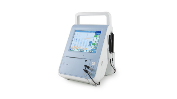
Biometer and Pachymeter
| Probe Frequency: | 10Mhz; 20Mhz |
|---|---|
| Resolution: | 0.01mm; 1um |
| Accuracy: | ±0.1mm; ±5um |
| Measurement Range: | 15~35mm(vel 1640m/s); 0.23~1.3mm |
| Gain adjustable: | 0-120dB |
| Measurements: | ACD, Lens, Vitreous and AXL |
| Power Requirement: | AC 100-240V 50-60Hz|Rate power:≤45VA |
| Dimension: | 200*168*165mm |
OD1-A/P/AP Ophthalmological Ultrasonic Measuring Instruments
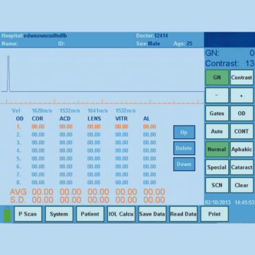
Nearly 20 years ultrasound research, OD1 series for ophthalmology biometry is our classical gift to doctors. With 5.7” color Led touch screen, sensitive and accurate measurement, it is a single device, but it can realize three models of applications.
- Product Parameter
Biometer
- Probe Frequency: 10 Mhz
Resolution: 0.01mm
Accuracy: ±0.1mm
Gain adjustable: 0-120dB
Measurement range: 15~35 mm (vel 1640m/s)
Measurements: ACD, Lens, Vitreous, and AXL
Examination modes: normal/aphakia/dense cataract/PMMA, Acrylic & Silicone.
Measurement mode: immersion and contact , auto and manual
IOL formulas: SRK-II,SRK-T,BINKHORST,HOLLADAY,HOFFER-Q,HAIGIS
Pachymeter
- Probe frequency: 20 Mhz
Resolution: 1um
Measurement range: 0.23~1.3mm
Accuracy: ±5um
Biometer/ A Scan
OD1-A Biomerter:
- Main unit
- 10.0 MHz A probe
- Gage block for A-scan/ Biometer
- Power adaptor
- Touch screen pen


Pachymeter/P Scan
OD1-P Pachymeter
- Main unit
- 20.0MHz angled probe
- Gage block for Pachymeter
- Power adaptor
- Touch screen pen
Bio & Pachymeter/ AP Scan
OD1-AP Biometer/Pachymeter
- Main unit
- 10.0MHz A probe
- 20.0MHz angled probe
- Two kinds of Gage blocks
- Power adaptor
- Touch screen pen

Related Products
What is A Type Ultrasound?
A Type Ultrasound is based on the relationship between the time and amplitude of the sound wave to detect the echo of the sound wave, and its positioning accuracy is high.
Ocular A type ultrasound is to place the probe in front of the eye, the sound beam propagates forward, and the echo is reflected once at each interface. The echo is arranged on the baseline in the form of A crest according to the return time, and the height of the crest indicates the echo intensity. The stronger the echo, the higher the crest.
It is difficult to interpret lesions by forming one-dimensional images with A-ultrasound, but it has high tissue discrimination. A super axial resolution is high, the liquid crystal digital display of anterior chamber depth, crystal thickness, vitreous cavity length and shaft length, accuracy up to 0.01mm, for the measurement of eye structure.
The A super keratometer can be used to measure corneal thickness with an accuracy of 0.01mm and is used to measure corneal thickness before refractive surgery.
The posterior optic nerve and ocular muscle could not be measured by A ultrasound.
At present, many A-ultrasounds have entered the intraocular lens calculation formula, and when the eye axis and corneal curvature are measured, the intraocular lens calculation mode can be automatically transferred to obtain the exact degree of the required intraocular lens.


