


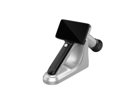


Portable Non-mydriatic Fundus Camera PFC11
This is a portable and smart fundus camera for rapid screening of fundus lesions.
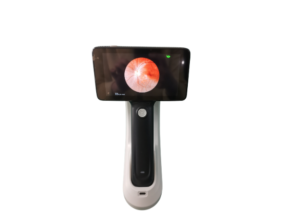
- Non-mydriatic Camera for Minimum 2.5mm Pupil Size
- High-definition Fundus Imaging
- Multi-point Target Fixation
- Auto and Manual Focus Mode
- AI Diagnosis Support
New Model Fundus Camera PFC11
This is Maxter second-generation Fundus imaging system. The model PFC11 fundus camera effectively reduces image noise, making the details of fundus image more realistic and much clearer.
Dados Técnicos
| Tipo | Non-Mydriatic/Portable |
| Fov | 50° |
| Sensor Resolution | 18 Mega pixel |
| Min. Pupil Diameter | 2.5milímetros |
| Fonte de luz | White LED &Infrared LED |
| Focus | Auto/Manual Focus |
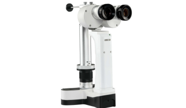
One Step To Be Table Fundus Camera

Type A Stand for Fundus Camera
This is our advanced stand designed for our portable fundus camera. It is also portable and can be taken with a small case.
Type B Stand for Fundus Camera
This is a valued fundus camera stand to make the photographing more stable and reliable. It is an affordable choice.

Other Technical Parameter
- Ficha de dados
| Screen | 4.7-inch Multi-touch Screen(Anti-glare) |
| Memory | 4G+64G |
| Data Transmission | USB3.0,WIFI, Bluetooth, SD, Type C |
| Other Functions | 4G, NFC |
| Bateria | 18650 Bateria recarregável |
| Peso líquido | <600g |


Package For Portable Fundus Camera

Fundo Imagens


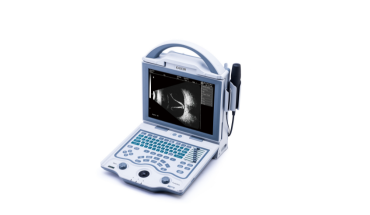

Fundus camera is a special device used to take human fundus images, also known as fundus camera, or fundus photography equipment.
It has features such as high-resolution images, autofocus, lighting system, digital image processing, etc., which can help doctors observe and diagnose eye diseases.
The components are camera body, lens system, flash/ lighting system, stand or support system, digital image processing system, display screen and user interface.
Fundus cameras use optical principles and high-resolution imaging techniques to photograph the internal structure of the eye. It focuses light through a lens system, and sensors or cameras capture light that reflects or passes through the eye tissue to form an image. The lighting system ensures adequate light exposure, while the digital image processing system processes, stores and transmits images for doctors to analyze. These components work together to enable medical fundus cameras to provide clear, detailed images of eye structures for diagnosis and monitoring of eye health.
Diagnosis of retinal disease, screening for diabetic retinopathy, evaluation of glaucoma, evaluation of fundus vascular disease, evaluation of cataract surgery, diagnosis of ocular inflammation and infection, etc..